The Mammalian Placenta and Early Mammalian Development
The Mammalian Placenta
and Early Mammalian
Development
Because mammals are descendants of
one of three lineages that originated
with a common amniote ancestor, they
inherited the amniotic egg. However,
rather than developing within the egg
shell like other amniotes, most mammalian
embryos evolved the propitious
strategy of developing within the
mother’s body. We have already seen
that mammalian gastrulation closely
parallels that of the egg-laying amniotes.
The earliest mammals were egg
layers, and even today some mammals
retain this primitive character; the monotremes (duck-billed platypus
and spiny anteater) lay large yolky
eggs that closely resemble bird eggs. In marsupials (pouched mammals such
as opossums and kangaroos), the
embryos develop for a time within the
mother’s uterus. But the embryo does
not “take root” in the uterine wall, and
consequently it receives little nourishment
from the mother before birth.
The young of marsupials are therefore
born immature and are sheltered in a
pouch in the mother’s abdominal wall
and nourished with milk.
All other mammals, composing 94% of class Mammalia, are placental mammals. These mammals have evolved a placenta, a remarkable fetal structure through which the embryo is nourished. Evolution of this fetal organ required substantial restructuring, not only of the extraembryonic membranes to form the placenta but also of the maternal oviduct, part of which had to expand into long-term housing for the embryo, the uterus. Despite these modifications, development of the extraembryonic membranes in placental mammals is remarkably similar to their development in egg-laying amniotes (compare Figures 8-20 and 8-22).
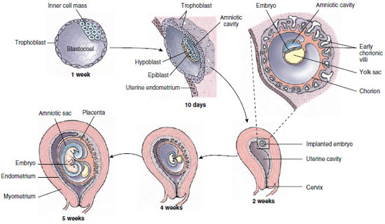
The early stages of mammalian
cleavage, shown in Figure 8-7E, occur
while the blastocyst is traveling down
the oviduct toward the uterus, propelled
by ciliary action and muscular
peristalsis. When a human blastocyst is
about six days old and composed of
about 100 cells, it contacts the uterine
endometrium (uterine lining) (Figure
8-23). On contact, the trophoblast
cells proliferate rapidly and produce
enzymes that break down the epithelium
of the uterine endometrium.
These changes allow the blastocyst to
implant in the endometrium. By the
eleventh or twelfth day the blastocyst
is completely buried and surrounded
by a pool of maternal blood. The trophoblast
thickens, sending out thousands
of tiny, fingerlike projections,
the chorionic villi. These projections
sink like roots into the uterine endometrium
after the embryo implants. As
development proceeds and embryonic
demands for nutrients and gas exchange
increase, the great proliferation of chorionic
villi vastly increases the total surface
area of the placenta. Although the
human placenta at term measures only
18 cm (7 inches) across, its total absorbing
surface is approximately 13 square
meters—50 times the surface area of the
skin of the newborn infant.
One of the most intriguing questions the placenta presents is,why is it not immunologically rejected by the mother? Both placenta and embryo are genetically alien to the mother because they contain proteins (called major histocompatibility proteins) that differ from those of the mother.We would expect uterine tissues to reject the embryo just as the mother would reject an organ transplanted from her own child.The placenta is a uniquely successful foreign transplant, or allograft, because it has evolved measures for suppressing the immune response that normally would be mounted against it and the fetus by the mother.Recent experiments suggest that the chorion produces proteins and lymphocytes that block the normal immune response by suppressing formation of specific antibodies by the mother.
Since the mammalian embryo is protected and nourished through the placenta rather than with stored yolk, what becomes of the four extraembryonic membranes it has inherited from the early amniotes? The amnion remains unchanged, a protective water jacket in which the embryo floats. A fluid-filled yolk sac is also retained, although it contains no yolk. It has acquired a new function: during early development it is the source of stem cells that give rise to blood and lymphoid cells. These stem cells later migrate into the developing embryo. The two remaining extraembryonic membranes, the allantois and the chorion, are recommitted to new functions. The allantois is no longer needed for the storage of metabolic wastes. Instead it contributes to the umbilical cord, which links the embryo physically and functionally with the placenta. The chorion, the outermost membrane, forms most of the placenta itself. The rest of the placenta is formed by the adjacent uterine endometrium.
The embryo grows rapidly, and all of the major organs of the body have begun their formation by the end of the fourth week of development. The embryo is now about 5 mm in length and weighs approximately 0.02 g. During the first two weeks of development (the germinal period) the embryo is quite resistant to outside influences. However, during the next eight weeks, when all of the major organs are being established and body shape is forming (the embryonic period), the embryo is more sensitive to disturbances that might cause malformations (such as exposure to alcohol or drugs taken by the mother) than at any other time in its development. The embryo becomes a fetus at approximately two months after fertilization. This ushers in the fetal period, which is primarily a growth phase, although some of the organ systems (especially the nervous and endocrine systems) will continue to develop. The fetus grows from approximately 28 mm and 2.7 g at 60 days to approximately 350 mm and 3000 g at term (nine months).
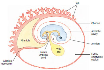 |
| Figure 8-22 Generalized diagram of the extraembryonic membranes of a mammal, showing how their development parallels that of a chick (compare with Figure 8-20). Most extraembryonic membranes of the mammal have been redirected to new functions. |
All other mammals, composing 94% of class Mammalia, are placental mammals. These mammals have evolved a placenta, a remarkable fetal structure through which the embryo is nourished. Evolution of this fetal organ required substantial restructuring, not only of the extraembryonic membranes to form the placenta but also of the maternal oviduct, part of which had to expand into long-term housing for the embryo, the uterus. Despite these modifications, development of the extraembryonic membranes in placental mammals is remarkably similar to their development in egg-laying amniotes (compare Figures 8-20 and 8-22).
|
One of the most intriguing questions the placenta presents is,why is it not immunologically rejected by the mother? Both placenta and embryo are genetically alien to the mother because they contain proteins (called major histocompatibility proteins) that differ from those of the mother.We would expect uterine tissues to reject the embryo just as the mother would reject an organ transplanted from her own child.The placenta is a uniquely successful foreign transplant, or allograft, because it has evolved measures for suppressing the immune response that normally would be mounted against it and the fetus by the mother.Recent experiments suggest that the chorion produces proteins and lymphocytes that block the normal immune response by suppressing formation of specific antibodies by the mother.
Since the mammalian embryo is protected and nourished through the placenta rather than with stored yolk, what becomes of the four extraembryonic membranes it has inherited from the early amniotes? The amnion remains unchanged, a protective water jacket in which the embryo floats. A fluid-filled yolk sac is also retained, although it contains no yolk. It has acquired a new function: during early development it is the source of stem cells that give rise to blood and lymphoid cells. These stem cells later migrate into the developing embryo. The two remaining extraembryonic membranes, the allantois and the chorion, are recommitted to new functions. The allantois is no longer needed for the storage of metabolic wastes. Instead it contributes to the umbilical cord, which links the embryo physically and functionally with the placenta. The chorion, the outermost membrane, forms most of the placenta itself. The rest of the placenta is formed by the adjacent uterine endometrium.
The embryo grows rapidly, and all of the major organs of the body have begun their formation by the end of the fourth week of development. The embryo is now about 5 mm in length and weighs approximately 0.02 g. During the first two weeks of development (the germinal period) the embryo is quite resistant to outside influences. However, during the next eight weeks, when all of the major organs are being established and body shape is forming (the embryonic period), the embryo is more sensitive to disturbances that might cause malformations (such as exposure to alcohol or drugs taken by the mother) than at any other time in its development. The embryo becomes a fetus at approximately two months after fertilization. This ushers in the fetal period, which is primarily a growth phase, although some of the organ systems (especially the nervous and endocrine systems) will continue to develop. The fetus grows from approximately 28 mm and 2.7 g at 60 days to approximately 350 mm and 3000 g at term (nine months).
Support our developers


