Gametogenesis
Gametogenesis
The series of transformations that results in the formation of mature gametes is called gametogenesis. Although the same essential processes are involved in the maturation of both sperm and eggs, there are some important differences. Gametogenesis in testes is called spermatogenesis, and in ovaries, oogenesis.
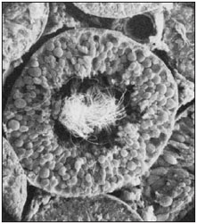
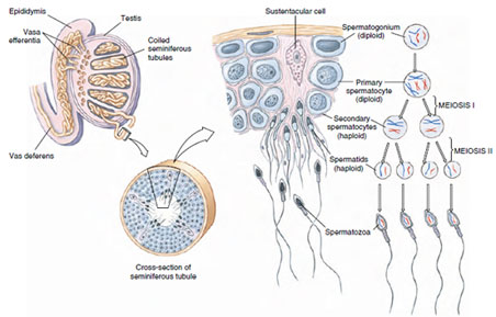
Spermatogenesis The walls of the seminiferous tubules contain differentiating germ cells arranged in a stratified layer five to eight cells deep (Figure 7-7). Germ cells develop in close contact with large sustentacular (Sertoli) cells, which extend from the periphery of the seminiferous tubules to the lumen and provide nourishment during germ cell development and differentiation (Figure 7-8). The outermost layers contain spermatogonia, diploid cells that have increased in number by ordinary mitosis. Each spermatogonium increases in size and becomes a primary spermatocyte. Each primary spermatocyte then undergoes the first meiotic division, as described previously, to become two secondary spermatocytes.
Each secondary spermatocyte enters the second meiotic division without the intervention of a resting period. In the two steps of meiosis each primary spermatocyte gives rise to four spermatids, each containing the haploid number (23 in humans) of chromosomes. A spermatid may contain all chromosomes that the male inherited from his mother, those he inherited from his father, or most likely, a combination of his parents’ chromosomes. Without further divisions the spermatids are transformed into mature spermatozoa or (sperm) (Figure 7-8). Modifications include great reduction of cytoplasm, condensation of the nucleus into a head, formation of a middle piece containing mitochondria, and a whiplike, flagellar tail for locomotion (Figure 7-8, 7-9). The head consists of a nucleus containing the chromosomes for heredity and an acrosome, a distinctive feature of nearly all the metazoa (exceptions are teleost fishes and certain invertebrates). In many species, both invertebrate and vertebrate, the acrosome contains lysins that serve to clear an entrance through the layers that surround the egg. In mammals at least, one of the lysins is the enzyme hyaluronidase, which allows the sperm to penetrate the follicular cells surrounding the egg. A striking feature of many invertebrate spermatozoa is the acrosome filament, an extension of varying length in different species that projects suddenly from the sperm head when the latter first contacts the surface of the egg. The fusion of the egg and sperm plasma membranes is the initial event of fertilization (See Contact and Recognition between Egg and Sperm).
The total length of a human sperm is 50 to 70 µm. Some toads have sperm that exceed 2 mm (2000 µm) in length (Figure 7-9) and are easily visible to the unaided eye. Most sperm, however, are microscopic in size. In all sexually reproducing animals the number of sperm in males is far greater than the number of eggs in corresponding females. The number of eggs produced is related to the chances of the young to hatch and reach maturity.
The series of transformations that results in the formation of mature gametes is called gametogenesis. Although the same essential processes are involved in the maturation of both sperm and eggs, there are some important differences. Gametogenesis in testes is called spermatogenesis, and in ovaries, oogenesis.
Spermatogenesis The walls of the seminiferous tubules contain differentiating germ cells arranged in a stratified layer five to eight cells deep (Figure 7-7). Germ cells develop in close contact with large sustentacular (Sertoli) cells, which extend from the periphery of the seminiferous tubules to the lumen and provide nourishment during germ cell development and differentiation (Figure 7-8). The outermost layers contain spermatogonia, diploid cells that have increased in number by ordinary mitosis. Each spermatogonium increases in size and becomes a primary spermatocyte. Each primary spermatocyte then undergoes the first meiotic division, as described previously, to become two secondary spermatocytes.
Each secondary spermatocyte enters the second meiotic division without the intervention of a resting period. In the two steps of meiosis each primary spermatocyte gives rise to four spermatids, each containing the haploid number (23 in humans) of chromosomes. A spermatid may contain all chromosomes that the male inherited from his mother, those he inherited from his father, or most likely, a combination of his parents’ chromosomes. Without further divisions the spermatids are transformed into mature spermatozoa or (sperm) (Figure 7-8). Modifications include great reduction of cytoplasm, condensation of the nucleus into a head, formation of a middle piece containing mitochondria, and a whiplike, flagellar tail for locomotion (Figure 7-8, 7-9). The head consists of a nucleus containing the chromosomes for heredity and an acrosome, a distinctive feature of nearly all the metazoa (exceptions are teleost fishes and certain invertebrates). In many species, both invertebrate and vertebrate, the acrosome contains lysins that serve to clear an entrance through the layers that surround the egg. In mammals at least, one of the lysins is the enzyme hyaluronidase, which allows the sperm to penetrate the follicular cells surrounding the egg. A striking feature of many invertebrate spermatozoa is the acrosome filament, an extension of varying length in different species that projects suddenly from the sperm head when the latter first contacts the surface of the egg. The fusion of the egg and sperm plasma membranes is the initial event of fertilization (See Contact and Recognition between Egg and Sperm).
|
|
The total length of a human sperm is 50 to 70 µm. Some toads have sperm that exceed 2 mm (2000 µm) in length (Figure 7-9) and are easily visible to the unaided eye. Most sperm, however, are microscopic in size. In all sexually reproducing animals the number of sperm in males is far greater than the number of eggs in corresponding females. The number of eggs produced is related to the chances of the young to hatch and reach maturity.
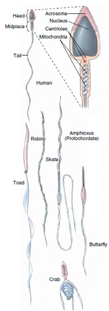 |
| Figure 7-9 Types of vertebrate and invertebrate sperm. |
Support our developers


